
Table of Contents
While researching transcranial magnetic stimulation (TMS), you will realize that there are various techniques for performing the treatment. The two most common techniques for mapping a target location nare scalp-based and MRI-guided treatments. In this blog post, we will discuss the two major techniques and the differences and benefits between them.
What is the 10-20 system?
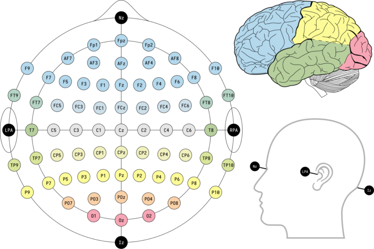
The 10-20 system is used to map the entire skull for general TMS by measuring specific percentages between skull points. The calculation is often done automatically by the 3D mapping software.
Four spots are used for the landmarks:
- Nasion or the bridge of the nose
- Top of the tragus of the left and right ears
- Top of the scalp
To target the depression site, or the left dorsolateral prefrontal cortex (LDLPFC), the clinician locates the motor cortex, also known as C3, and then measures 5 cm forward to approximate the treatment site, F3.
What is the Beam F3 method?
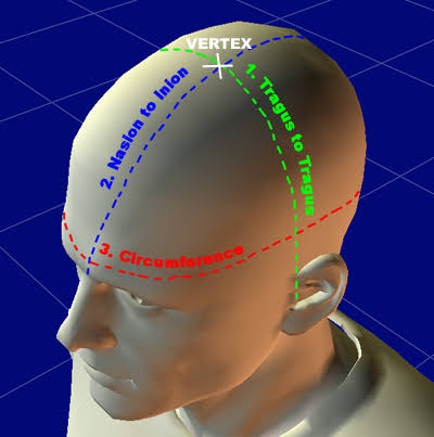
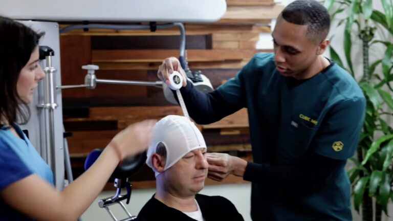
The Beam-F3 method uses a flexible tape measure to find three measurements:
- Tragus-to-tragus distance
- Nasion-to-ionion (bump in the back of the head) distance
- Head circumference
These three numbers are entered into a formula that calculates the exact placement coordinates, which are then marked on the cap.
A disadvantage of this method is that if the cap moves or is not placed in exactly the same position each time, the placements will be slightly different between treatments. NeoPsych does not use caps; however, this can be accommodated upon request.
What is 3-D-Guided Scalp-Based TMS?
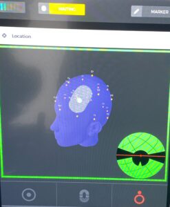
Using the navigation software in the MagStim device, anatomical landmarks are marked in 3D space. Scalp-based TMS uses the 10-20 system to automatically determine the treatment location with the device’s software. Once the anatomical landmarks of the ears, the bridge of the nose, and the top of the scalp are identified, the motor threshold location on the motor cortex is determined using the C3-marked area in the software after the head measurement is taken. Then, the F3 or the LDLPFC treatment site is found 5 cm from the C3 location. This is the treatment spot used for all TMS treatments.
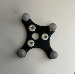
Unlike the Beam F3 method, 3-D-guided scalp-based TMS does not use a cap. Instead, a head reference is used to determine whether there are any movements from the treatment location, thereby providing greater precision in the treatment spot.
What is MRI-Guided TMS?
Using a functional MRI, the treatment location is picked in the left dorsolateral prefrontal cortex. The chosen spot has heightened activity in the brain. The area of heightened activity is chosen because it is anti-correlated with the subgenual anterior cingulate cortex. This area is chosen based on a neuroscientist’s analysis.
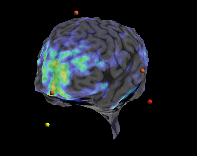
The subgenual anterior cingulate cortex serves as the bridge between our forebrain and the brain’s emotional center, the amygdala.
Thus, MRI tends to provide the most accurate localization, as it is tailored to each patient’s brain and prefrontal cortex activity. If you are looking for the most customizable and personalized treatment location, MRI-guided TMS is the best option.
The con of MRI-guided treatment is the time and money required to obtain an MRI.
Does Insurance Cover MRI-guided TMS?
While many insurance plans cover TMS for FDA-cleared indications when eligibility requirements are met, additional costs associated with MRI-guided neuronavigation may not always be covered. Patients should verify benefits with both their provider and insurance company.
Is MRI-Guided TMS More Effective?
MRI-guided neuronavigation offers greater precision in determining the treatment location, as the scan identifies a specific site based on brain activity. However, this does not directly translate into improved clinical outcomes on its own, as other factors play into the effect, such as the stimulation protocol and the condition being treated.
Some studies suggest personalized targeting may improve consistency and potentially enhance outcomes for certain patients, while others have found similar symptom improvement with experienced clinicians using scalp-based methods.
Which Option Should I Choose?
The treatment technique you choose depends on personal preference and how personalized you want the treatment to be. Both techniques are used throughout TMS clinics and are effective. Considering your financial and time constraints is important when deciding which treatment method to choose. Our team at NeoPsych would be more than happy to discuss any questions you may have regarding the treatment method you are using.
As the prefrontal cortex strengthens its communication with the amygdala, emotional processing begins to change. During this adjustment period, some patients may temporarily experience heightened emotional sensitivity as the brain reaches a new balance.
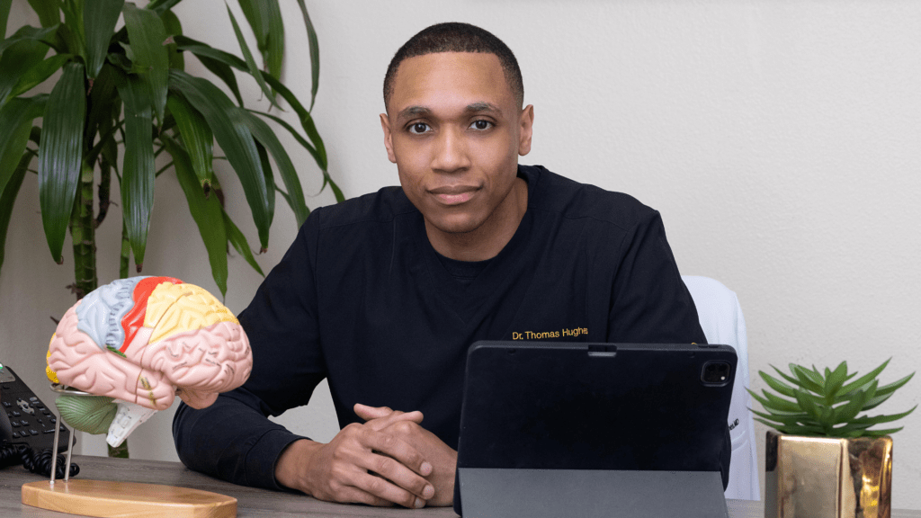
Dr. Thomas Hughes is an adolescent and adult interventional psychiatrist. He is the owner and founder of Heard Medical and NeoPsych in Glendale, CA. He specializes in medication management and Transcranial Magnetic Stimulation for depression, anxiety, OCD, and PTSD.
Esha Nushrat is a pre-medical student interested in Neurology. She has been working in mental health and volunteers at organizations such as Stride in Recovery and NAMI
This blog post has been medically reviewed by Dr. Thomas Hughes, the practicing provider at NeoPsych.