MRI-Guided TMS
In Glendale, Burbank & Los Angeles
M-SAINT TMS (fcMRI) vs. SAINT TMS
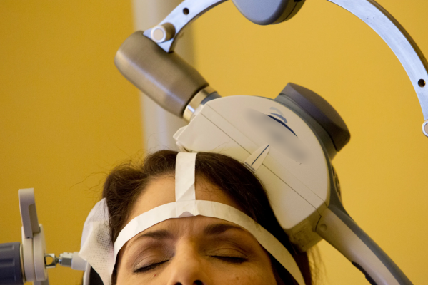
The benefit of TMS is that you only need to stimulate the very surface of the brain in order to send signals into the deeper areas that control depression and anxiety. Functional connectivity is able to maximize the location to which these signals are sent.
For the purposes of TMS, the goal is to identify the target area most connected to the subgenual anterior cingulate cortex (sgACC). Studies have shown that the sgACC is dysregulated in patients suffering from depression. TMS seeks to correct this dysregulation. Without post-processing an fMRI, it is impossible to identify the location on the brain that maximizes access to the sgACC. Once discovered, your TMS provider will determine how to adjust the activity of the sgACC for the purposes of treatment.
The differences between fcMRI and SAINT protocol are that SAINT uses a specific machine and specific algorithm to locate the sgACC connectivity. Please watch the video below for more detail.
Of note, there is an anti-correlation between DLPFC and sgACC. (reference) i.e. For depression, there is typically less activity in the DLPFC (dorsolateral prefrontal cortex) and too much activity in the sgACC. The goal of the post processing is to locate where this anti-correlation is most active.
Functional MRI vs. Functional Connectivity MRI
Functional MRIs are able to detect the movement of blood throughout the body. It can do this because the oxygen carried via blood has a magnetic field that changes as it is being used. If there is a change in magnetic field, an MRI can take a picture of it. The more the brain is working/functioning, the more oxygen molecules that can be detected by the MRI. Thus, we are able to see the most functional areas of the brain. (reference)
For the purposes of TMS, we can use fMRIs to locate the most functional areas of the brain for treating conditions like Depression, Anxiety, and OCD. Unfortunately, a functional MRI is just another MRI unless it is analyzed by a professional to see how and where the most functional areas are connected to other areas of the brain. The analysis results in a functional connectivity MRI, which is very important for customizing TMS treatment.

What is an MRI? Do You Need It For Accelerated TMS?
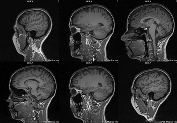
For the purposes of TMS, we can use an individual’s MRI to identify specific target locations on their brain to administer customized treatment.
MRI stands for Magnetic Resonance Imaging. This is a method of taking pictures of the internal structures of the body. Unlike an X-ray, which uses external radiation in order to take a picture, MRIs use a very powerful magnet. This technique reduces radiation exposure and also enhances the detail of the obtained images.
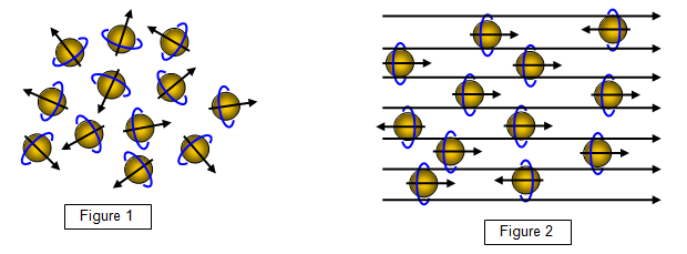
The magnet in an MRI is very powerful. The magnet can painlessly change the direction that the particles in your body point. When the particles in your body return to their initial direction, the MRI machine is able to use the movement to create a picture. (reference)
What if I don't want to get an MRI?
3D guided TMS
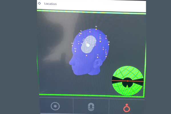
3DG TMS uses the traditional methods of mapping. It does not use an MRI. 3DG is a method of RECREATING the angle and location of the target from session to session. 3DG does this by creating an individualized head model of each patient. This model allows the provider to place the coil in the same location each day, maximizing the consistency between sessions. It also helps providers visualize if the treatment coil moves out of place during treatment. With 3DG there is no need for a cap or measuring the treatment location.
Traditional TMS (Cap/Measurements)
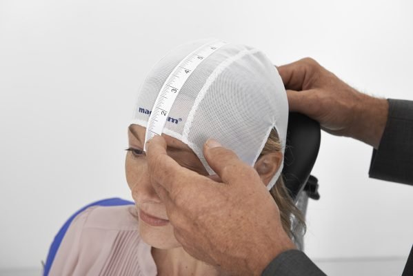
Using a cap and anatomical measurements require traditional methods of mapping and target location selection. Neither of these methods use MRI. These methods are perfectly reasonable and have been a trusted part of TMS since the procedure became available to the public in 2008. There are issues with consistency from session to session and head movement.
